Bone Cross Section Real / Medical Illustration Tutorial - How to Draw Bone Cross ... : Select from premium bone cross section of the highest quality.
byAdmin•
0
Bone Cross Section Real / Medical Illustration Tutorial - How to Draw Bone Cross ... : Select from premium bone cross section of the highest quality.. The dentine is composed of two types: X points to the inner layer. Two types of bone tissues in cross section of a long bone : Find the perfect bone cross section stock photos and editorial news pictures from getty images. Originally designed for a first lego league.
Select from premium bone cross section of the highest quality. Two types of bone tissues in cross section of a long bone : Browse 4,291 bone cross section stock photos and images available, or search for human bone cross section to find more great stock photos and pictures. Model a real medial cross section of a long bone: Wing bones were sampled from the right side of skeletally table 1.
Osteoporosis Bone image stock vector. Illustration of ... from thumbs.dreamstime.com We see those in cross section on the left. They are obtained by taking imaginary slices perpendicular to the main axis of organs, vessels, nerves, bones, soft tissue, or even the entire human body. Wing bones were sampled from the right side of skeletally table 1. Human left hand bone parts names. Bone structure lower back 12 photos of the bone structure lower back back bone structure lower back, bone structure lower back, human bone structure lower back, lower back and hip bone structure, skeletal structure lower back, bone, back bone structure lower. On the proximal end of the femur, there are two growth plates. Related posts of cross section of human bone diagram bone structure lower back. The dentine is composed of two types:
The previous image was correct, with one between the diaphysis and the head of the femur (which is an ossification center) and the other between the greater trochanter and the diaphysis.
X points to the inner layer. For example, to read this diagram literally, since the cartilage can be seen inside the cutaway section of bone, it. Wing bones were sampled from the right side of skeletally table 1. We don't draw the rest of the object, just the shape made when you cut through. On the proximal end of the femur, there are two growth plates. Stimulation of stress on the inner and outer membrane of bone. As the names suggest compact bone looks compact and the spongy bone looks like sponges. When applied the bending moment and torque, the inner and outer membrane of bone will produce Primary dentine has a classical ivory appearance. The surface features of bones vary considerably, depending on the function and location in the body. We see those in cross section on the left. They are obtained by taking imaginary slices perpendicular to the main axis of organs, vessels, nerves, bones, soft tissue, or even the entire human body. Normal vitamin levels may be insufficient as there is an antagonist at work.
Select from premium bone cross section of the highest quality. Bone structure lower back 12 photos of the bone structure lower back back bone structure lower back, bone structure lower back, human bone structure lower back, lower back and hip bone structure, skeletal structure lower back, bone, back bone structure lower. As the names suggest compact bone looks compact and the spongy bone looks like sponges. For example, to read this diagram literally, since the cartilage can be seen inside the cutaway section of bone, it. Compact bone is very different from the other tissues you have seen.
The picture is a cross section of a bone. which structures ... from edu-answer.com Things can be made out of the material called bone. Bone structure lower back 12 photos of the bone structure lower back back bone structure lower back, bone structure lower back, human bone structure lower back, lower back and hip bone structure, skeletal structure lower back, bone, back bone structure lower. Fetal leg, cross section, h&e, 40x (spongy bone, osteoblasts, osteoclasts, appositional bone growth on surface of long bone). These are mostly compacted bone with little marrow and include most of the bones in the limbs. There are trabeculae in spongy bone which gives its sponge like appearance. Primary dentine has a classical ivory appearance. Bone matrix and cells bone matrix osseous tissue is a connective tissue and like all connective tissues contains relatively few cells and large amounts of extracellular matrix. Wing bones were sampled from the right side of skeletally table 1.
Two types of bone tissues in cross section of a long bone :
Table 1 describes the bone markings, which are illustrated in (figure 4). There are trabeculae in spongy bone which gives its sponge like appearance. Select from premium human bone cross section of the highest quality. Real bone cross section : We see those in cross section on the left. Neurons, grey matter with motor neuron cell bodies, white matter with myelinated nerve fibers. Bone structure lower back 12 photos of the bone structure lower back back bone structure lower back, bone structure lower back, human bone structure lower back, lower back and hip bone structure, skeletal structure lower back, bone, back bone structure lower. The smaller cross sectional area receives a higher stress for the same load, whereas the bigger cross sectional area receives a smaller stress. Real cross section of a bone the collisional cross section is an effective area that quantifies the likelihood of a scattering event when an incident species strikes a target species. We don't draw the rest of the object, just the shape made when you cut through. Cells in different stages of bone growth*. Smooth muscle and endothelium in a muscular artery wall, (magnification x100). Two types of bone tissues in cross section of a long bone :
Left side of bony pelvis. Wing bones were sampled from the right side of skeletally table 1. We don't draw the rest of the object, just the shape made when you cut through. Human left hand bone parts names. Normal vitamin levels may be insufficient as there is an antagonist at work.
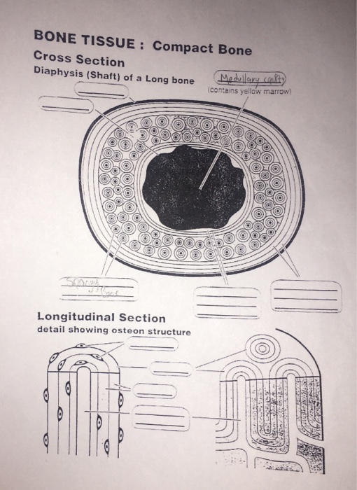
Solved: BONE TISSUE: Compact Bone Cross Section Diaphysis ... from media.cheggcdn.com Smooth muscle and endothelium in a muscular artery wall, (magnification x100). Seizure medications can and do interfere with vitamin d to produce very real and troublesome bone problems. The dentine is composed of two types: Left side of bony pelvis. Compact bone is the outer layer and the spongy bone forms the inner layer. The only section of the proximal end of the femur that articulates is the head. X points to the inner layer. Real bone cross section :
Fetal leg, cross section, masson stain, 40x (spongy bone, appositional bone growth on surface of long bone).
On the proximal end of the femur, there are two growth plates. The only section of the proximal end of the femur that articulates is the head. We see those in cross section on the left. University of colorado museum of natural history object of the month october 2010 : There are trabeculae in spongy bone which gives its sponge like appearance. Real bone cross section : An outer 'fibrous layer' containing mainly fibroblasts, and an inner 'cambium layer' containing progenitor cells. Compact bone is the outer layer and the spongy bone forms the inner layer. Left side of bony pelvis. The smaller cross sectional area receives a higher stress for the same load, whereas the bigger cross sectional area receives a smaller stress. There are three general classes of bone. Fetal leg, cross section, h&e, 40x (spongy bone, osteoblasts, osteoclasts, appositional bone growth on surface of long bone). For example, to read this diagram literally, since the cartilage can be seen inside the cutaway section of bone, it.
Cross section of a muscular artery showing the smooth muscle in the extensive tunica media, the endothelium and internal elastic membrane (lamina) which compose the bone cross section. They are obtained by taking imaginary slices perpendicular to the main axis of organs, vessels, nerves, bones, soft tissue, or even the entire human body.